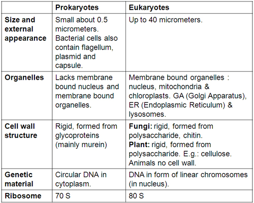
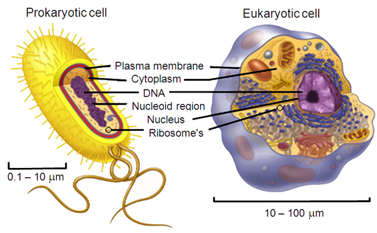
Types of cells:
-> Prokaryotic : Bacteria and Archaea (Sometimes Archaea are preferably classified altogether as a different class and not under Prokaryotes).
-> Eukaryotic : Unicellular (protists) and Multicellular (Fungi, plants and animals).
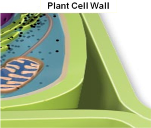
They provide protection, support, shape and rigifity to the cellular structure.
They also provides a porous medium for the circulation and distribution of water, minerals, and other small nutrient molecules.
They contains specialized molecules that regulate growth and protect the plant from disease.
Prokaryotes, especially bacteria are divided into Gram positive and Gram negative cells based on differential staining by crystal violet-iodine reagent (Grams reagent).
Bacteria stained purple are Gram + : their cell walls are thick and composed of predominantly petidoglycan and teichoic acid polymers which lipids are conspicuousy absent. The teichoic acid polymers play key role in antigenicity of the cell.
Bacteria stained pink are Gram : their cell walls have thin peptidoglycan (sandwiched between the cell membrane and outer envelope) and lipopolysaccharides with no teichoic acid.
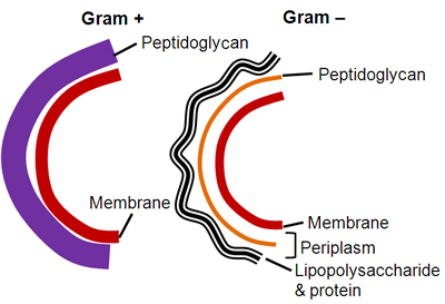
In Gram - negative bacteria, the outer membrane of lipopolysaccharides prevents the stain from reaching the peptidoglycan layer. The outer membrane is then permeabilized by acetone treatment, and the pink safranin counterstain is trapped by the peptidoglycan layer.